Sun–Mon: 9:00 AM – 9:00 PM · Tuesday Holiday

We excel in Women & Fetal Imaging
Fetal / Pregnancy Imaging
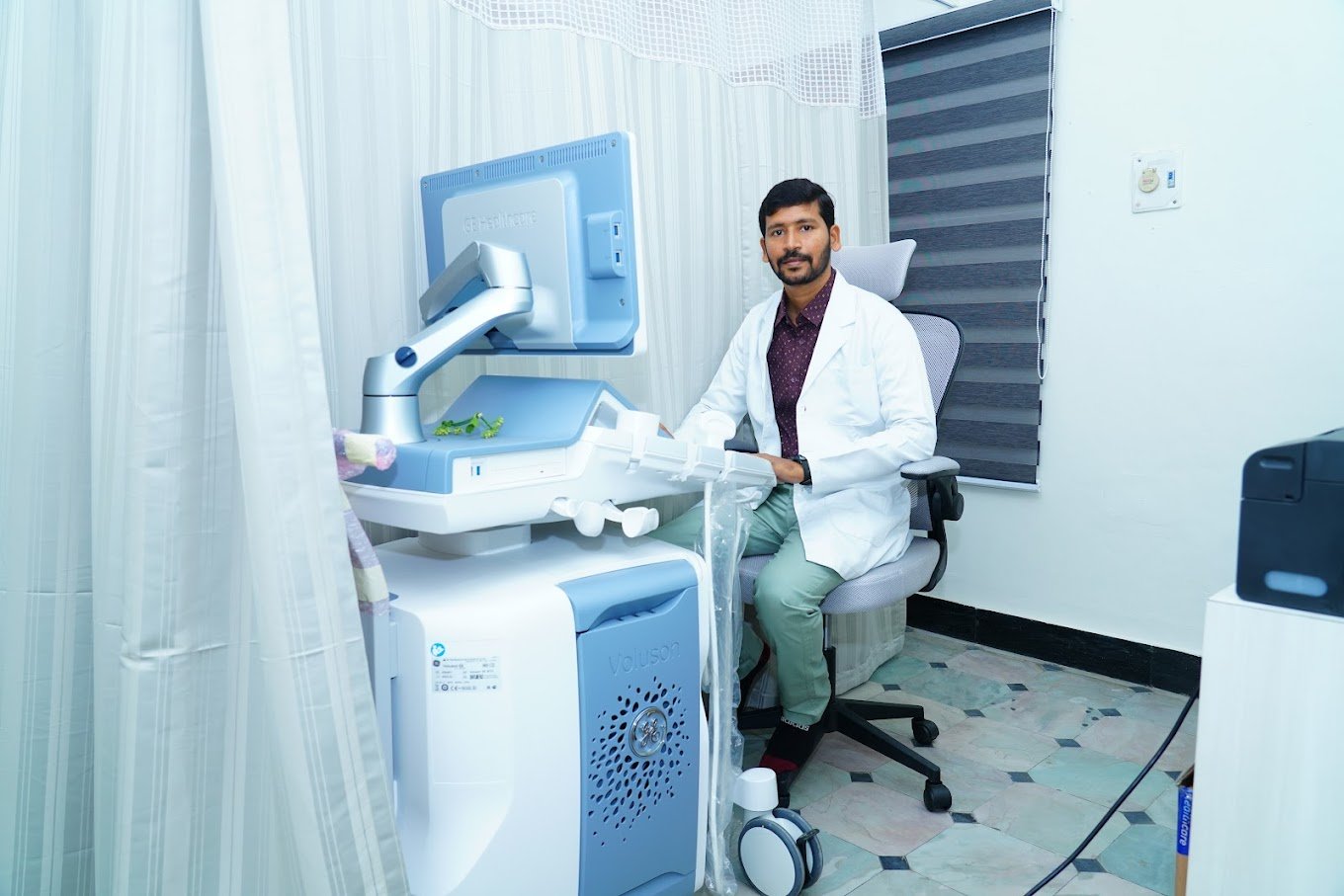
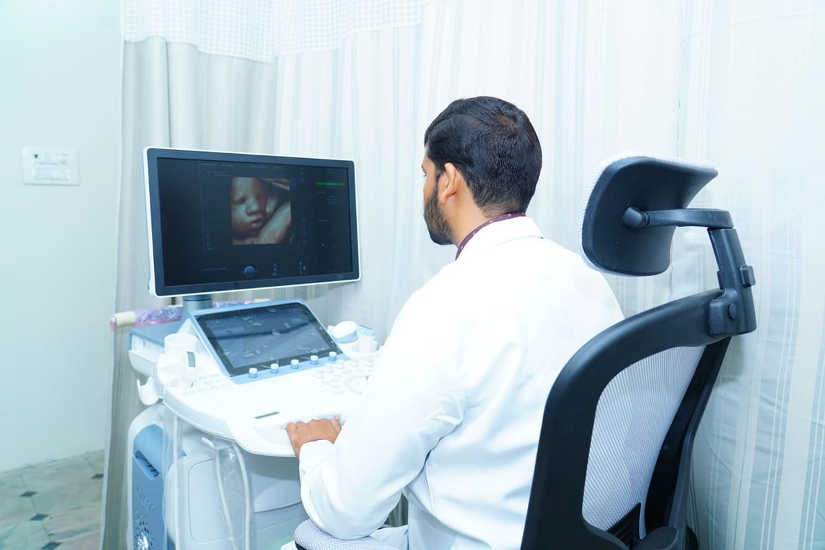
We monitor the health and development of your growing baby throughout pregnancy, offering comprehensive fetal/pregnancy imaging in Tirupati, including diagnostic pregnancy ultrasound and USG-guided interventions and invasive procedures.
- Pre pregnancy USG
- Early pregnancy ultrasound
- NT scan
- Early TIFFA scan
- TIFFA Scan
- Growth scan
- Fetal well being scan
- Doppler studies
- Post pregnancy USG
- Chorionic villus sampling
- Amniocentesis
- Fetal reduction procedures
- Cordocentesis
Women Imaging Services
Comprehensive women imaging in Tirupati, including infertility evaluation, follicular study, Sono HSG, PCOS evaluation, and general abdomen and pelvis ultrasound. Our experienced radiologists provide accurate and timely diagnoses for all women’s health concerns.
Our advanced imaging technology enables detailed evaluations of the reproductive system, including the uterus, ovaries, and fallopian tubes. We can identify potential causes of infertility, such as fibroids, polyps, or cysts.
Our follicular study imaging monitors the growth and development of ovarian follicles during the menstrual cycle, helping identify the best time for ovulation and increasing the chances of conception.
This specialized technique combines ultrasound and X-ray to provide a detailed evaluation of the uterus and fallopian tubes. It is a minimally invasive procedure that can identify potential blockages or abnormalities affecting fertility.
Polycystic Ovary Syndrome (PCOS) can cause hormonal imbalances and affect fertility. Our imaging can identify the characteristic cysts in the ovaries, as well as other issues such as thickened endometrial lining.
A comprehensive evaluation of the abdominal and pelvic regions, including the liver, gallbladder, pancreas, kidneys, bladder, uterus, and ovaries.
Mammography / X-Ray of Breast
Breast cancer is a leading cause of cancer deaths among women worldwide — but with early detection and timely treatment, the survival rate is high. We offer a range of advanced mammography and X-ray services to detect and diagnose breast abnormalities at an early stage.
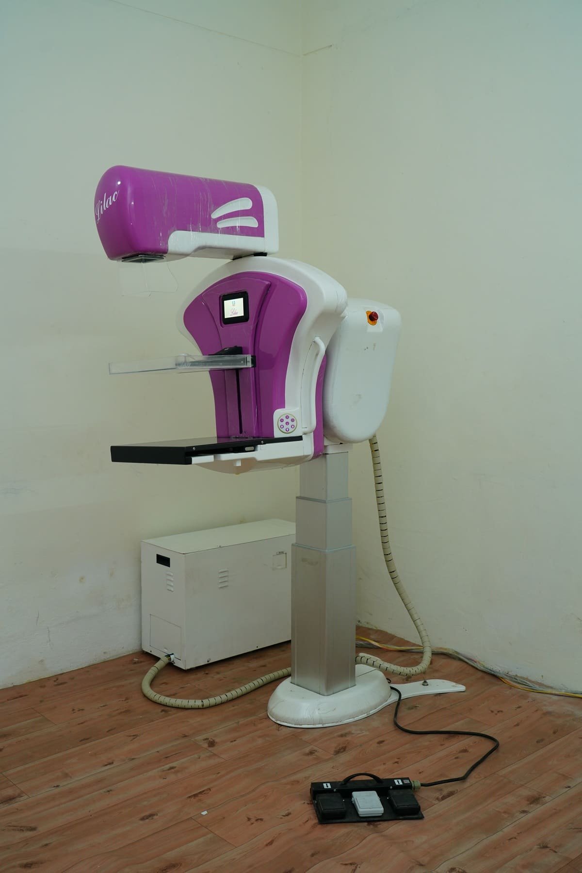
Low-dose X-rays examine the breast tissue to detect abnormalities and help find early-stage breast cancer before symptoms appear. Recommended for women over 40 or those with a family history of breast cancer.
High-frequency sound waves create detailed images of breast tissue. This non-invasive technique evaluates breast lumps or abnormalities and assesses breast health in women with dense breast tissue.
If a suspicious lump is detected, our team performs minimally invasive biopsy procedures, including ultrasound-guided and stereotactic biopsy, to obtain a tissue sample for analysis.
X-Ray / Radiography
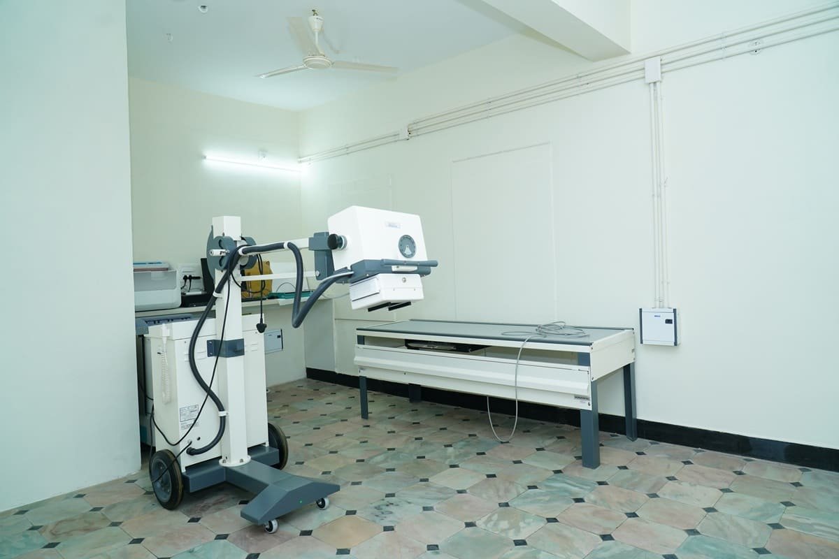
A comprehensive range of diagnostic imaging, including X-rays and radiography. These techniques use low levels of radiation to produce images of the body’s internal structures, allowing our radiologists to diagnose and monitor a range of conditions.
A special X-ray to evaluate the female reproductive system, particularly the uterus and fallopian tubes. A contrast dye is injected and X-rays assess its flow, helping diagnose conditions such as blocked fallopian tubes.
X-rays produce images of bones and internal organs, helping diagnose and monitor conditions such as fractures, arthritis, and pneumonia. Our state-of-the-art equipment ensures the highest quality images and care.
For suspicious findings on a mammogram or ultrasound, our team performs minimally invasive biopsy procedures to obtain a tissue sample for further analysis.
Schedule with ease and skip the wait
We understand that everyone has unique healthcare needs and that time is valuable. Our convenient online booking system ensures you won’t have to wait long for your scan.